ALLPCB


1. Introduction
Normally, the sinoatrial node in the right atrium generates electrical impulses automatically and rhythmically. These impulses are conducted through the cardiac conduction system to different parts of the heart, triggering myocardial contraction and pumping blood to the body. When the conduction system is impaired, or when the sinoatrial or atrioventricular nodes fail to generate or transmit regular impulses, arrhythmias or cardiac arrest can occur, threatening the patient's life. Cardiac pacemakers deliver direct electrical stimulation to the diseased heart as needed, artificially restoring a regular heartbeat. Pacemakers take two main forms: external temporary pacemakers for emergency temporary pacing, and implantable (permanent) pacemakers for long-term therapy. This article focuses on implantable pacemakers in the usual clinical sense.
2. Principles and Construction of a Pacemaker
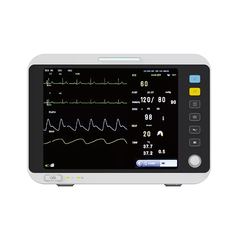
An implantable pacemaker is a compact, highly reliable electrical pulse generator. It consists of a pacing pulse generator connected to specialized leads (pacing catheter electrodes). The leads deliver pulses to the myocardium to pace the heart and also return sensed signals of intrinsic cardiac activity (intracardiac electrogram, ICG) to the pacemaker to control pulse delivery. A pacemaker is a medical electronic device.
An implantable pacemaker mainly consists of two parts:
(1) Pacing leads: they conduct the pacemaker's output signal to the myocardium for pacing and convey sensed intrinsic cardiac signals (intracardiac electrogram, ICG) back to the pacemaker to control pulse delivery. These endocardial electrodes have evolved from early unipolar designs to bipolar and even multipolar configurations. Leads intended for long-term pacing must be made of biocompatible, tough, aging-resistant, and corrosion-resistant materials. Lead conductors are typically made of Elgiloy or wound wires of nickel-chromium-cobalt-molybdenum alloy formed into a helical coil. Lead insulation commonly uses high-purity silicone rubber or medical-grade polyurethane. Electrode tips are often coated with activated isotropic low-temperature pyrolytic carbon or platinum.
(2) Pacing pulse generator: it comprises the pacing circuitry, battery, and metal housing. The power source must be compact, have high capacity, deliver energy slowly, be well sealed, and be reliable. Currently, implantable pacemakers worldwide commonly use lithium-iodine batteries, enabling continuous pacemaker service lives of over 10 years. Titanium is used for the hermetic housing because of its good biocompatibility and resistance to corrosion; housings are typically drawn and laser-welded with smooth rounded joints. Since the 1980s, pacing circuitry has commonly used integrated circuits. CMOS ASIC pacing chips, together with resistors, capacitors, reed switches, and other components, are mounted on ceramic substrates to form hybrid thick-film integrated circuits as standard pacing circuitry components.
3. Indications for Pacemaker Implantation
(1) High-grade or complete atrioventricular (AV) block accompanied by Adams-Stokes syndrome or syncope. Asymptomatic patients with heart rate <50 beats/min or wide QRS complex with ventricular pauses >2 seconds are relative indications.
(2) Complete or incomplete trifascicular or bifascicular block with intermittent or paroxysmal complete AV block, or ventricular rate <40 beats/min; bifascicular block with Adams-Stokes syndrome or syncope; alternating complete left and right bundle branch block with His bundle study confirming prolonged H-V interval.
(3) Second-degree type II AV block accompanied by Adams-Stokes syndrome or syncope. Persistent second-degree type II AV block or ventricular rate <50 beats/min without symptoms are relative indications.
(4) Sick sinus syndrome presenting as severe sinus bradycardia with ventricular rate <45 beats/min causing significantly reduced organ perfusion, angina, dizziness, or syncope; pronounced bradycardia, sinus arrest, or sinoatrial block with R-R intervals >2 seconds accompanied by syncope or Adams-Stokes attacks; bradycardia-tachycardia syndrome with syncope or Adams-Stokes attacks.
(5) Patients unresponsive to antiarrhythmic pacing, implantable cardioverter-defibrillators, or drug therapy for ectopic rapid arrhythmias.
(6) Recurrent carotid sinus syncope and episodes of ventricular asystole.
4. History and Current Status of Pacemakers
The widespread clinical use of pacemakers has enabled treatment of severe arrhythmias that were previously unresponsive to drugs, substantially reducing mortality from cardiovascular disease and representing a major contribution of modern biomedical engineering.
In 1932, thoracic surgeon Hyman in the United States invented the first spring-driven electrical pulse generator. Using two needle electrodes to puncture the atrium, he restored cardiac activity in cases of cardiac standstill and termed the device an artificial pacemaker, inaugurating the era of artificial pacing for arrhythmias.
Pacemakers were first used clinically in 1952, when Zoll in the United States used an external pacemaker applied to the chest wall to resuscitate two patients with life-threatening conduction block, accelerating clinical adoption. Implantable pacemakers were invented and applied clinically by Elmqvist in Sweden in 1958 and by Greatbatch in the United States in 1960. Since then, pacemakers evolved toward longer life, higher reliability, lighter weight, smaller size, and broader functionality.
Early pacemakers were fixed-rate (asynchronous) and suited mainly for permanent AV block and sick sinus syndrome; they were not suitable for intermittent bradycardia and could compete with intrinsic rhythm, sometimes worsening arrhythmias. In the mid-1960s, demand pacing modes appeared to allow synchronization with intrinsic activity. Atrial-triggered ventricular pacing (VAT) was developed for AV block, while ventricular inhibited pacing (VVI) became the most commonly used mode worldwide. In the 1970s, dual-chamber pacemakers that preserved AV sequence (DVI) and universal pacing modes capable of treating a variety of bradyarrhythmias (DDD) were introduced, completing the basic therapeutic functions of pacemakers.
In the 1980s, besides miniaturization, programmable and telemetry features were added. External programmers allowed adjustment of pacing mode, rate, amplitude, pulse width, sensing sensitivity, refractory period, and AV delay, and permitted telemetry of device status, battery use, myocardial impedance, patient data, and intracardiac electrograms to an external receiver for display. In the 1990s, developments included antitachycardia pacing and rate-adaptive pacing (DDDR), improving response to activity and treatment of lethal arrhythmias. More recently, higher-performance devices have emerged, including biventricular or atrioventricular-synchronized three-chamber pacemakers and devices combining pacing and defibrillation functions.
5. Pacemaker Coding
The North American Society of Pacing and Electrophysiology (NASPE) and the British Pacing and Electrophysiology Group (BPEG) use the NASPE/BPEG (NBG) code for device identification.
Typically, the first three code positions identify the paced chamber(s), the sensed chamber(s), and the response to sensing (P or R wave, or both). The optional fourth position denotes either programmability or rate responsiveness. P indicates one or two simple programmable functions; M denotes multiple programmable functions including mode, refractory period, sensing sensitivity, and pulse width. C indicates communication or rate-adaptive pacing based on one or more physiologic measurements. The fifth position denotes special antitachycardia features: P for antitachycardia pacing, S for cardioversion or defibrillation shock, D for dual function (pacing and shock). O indicates that the category or function is not provided.
Table 1: NASPE/BPEG (NBG) pacemaker code
6. Future Prospects
After decades of development, implantable pacemakers are now used beyond treating conduction block and brady-tachy syndromes; they are being explored for additional indications such as orthostatic hypotension, malignant neurocardiogenic syncope (vasovagal), congenital long QT syndrome, and isolated PR interval prolongation. These may represent future application directions.
Pacing circuitry centered on low-power microprocessor chips will allow device behavior to be modified in software, reducing reliance on full-custom ASIC designs and shortening development cycles and costs for new pacing circuitry.
With advances in computing and telemetry, fully automated pacemakers may become feasible. Such devices could adapt automatically to a patient’s electrophysiology, using time-series analysis, sensor inputs, and automatic interpretation of stored data to determine optimal pacing strategies. Pacemakers could automatically measure atrial and ventricular pacing and sensing thresholds and adjust baseline rates programmatically based on sensor data.