ALLPCB


Introduction
Compound action potentials of a nerve trunk are an objective sign of nerve excitation. Applying a stimulus of sufficient intensity to an excitable nerve trunk produces action potentials. The extracellular potential at the excited site becomes more negative than at the resting site. After the nerve impulse passes, the extracellular potential at the excited site returns to the resting level. This change in membrane potential during nerve trunk excitation is called the nerve trunk action potential.
If two recording electrodes are placed on the surface of an intact nerve trunk, excitation at one end will produce a propagated excitation wave that passes the two electrodes in sequence, producing two oppositely directed deflections called a biphasic action potential. If the nerve tissue between the two recording electrodes is disrupted so the excitation wave passes only the first electrode and cannot reach the second, only one-directional deflection is recorded, called a monophasic action potential.
A nerve trunk contains many nerve fibers, so the compound action potential differs from a single-fiber action potential: it is the summation of many individual fiber action potentials. In addition, the recording here is extracellular, which differs from intracellular recordings. Therefore, within a certain range the amplitude of the compound action potential can vary with stimulus intensity.
Conduction of action potentials along nerve fibers occurs at a finite velocity. Different fiber types have different conduction velocities, and thicker fibers conduct faster. For example, A-type fibers in amphibian sciatic nerve trunks conduct at about 35–40 m/s. Measuring the distance s that the nerve impulse travels and the time t required to traverse that distance yields the conduction velocity.
This experiment uses the ZL-620U medical signal acquisition and processing system to demonstrate how to record compound action potentials from nerve trunks, measure action potential parameters, and determine nerve conduction velocity.
Experimental Subjects and Materials
Subjects: Toads or frogs.
Equipment: ZL-620U medical signal acquisition and processing system, surgical instruments for amphibians, specimen shield box, several electrode leads, Ringer solution.
Methods and Procedure
1. Preparation of the sciatic nerve trunk specimen
Method 1: The specimen preparation is similar to that for a sciatic-gastrocnemius preparation.
Notes:
- The nerve trunk should be dissected as long as possible, from near the vertebrae proximally to the region near the ankle where the common peroneal and tibial nerves divide.
- Do not damage the nerve tissue during dissection, as this will affect the experiment.
- Tie fine threads to both ends of the nerve trunk and immerse the specimen in Ringer solution until use.
Method 2: Use the skinned lower trunk of the amphibian. Place the specimen prone, lift the distal sacral bone slightly with forceps, and remove the sacrum with scissors. Position the specimen supine, separate the sciatic nerves beside the vertebral column with a glass probe, thread and ligate the nerves near the vertebrae, and cut the nerves. Pull the suture ends dorsally through the sacral incision and pin the specimen prone. Gently lift the ligated nerve end to identify the sciatic nerve path, position scissors close to the femur and popliteal fossa, and cut the nerve with attached muscle along the nerve path down to the Achilles region. Cut the Achilles tendon and nerve. Grasp the excised nerve-muscle specimen and remove muscle tissue by gentle traction along the nerve trunk to obtain an isolated sciatic nerve trunk specimen.
2. Connect the experimental setup
1) Use leads to connect the instruments as shown in Figure 1. Connect the stimulus output of the ZL-620U to a pair of stimulating electrodes. Connect two pairs of recording electrodes to CH2 and CH4 of the ZL-620U. Connect the ground lead to earth. Avoid incorrect connections or poor contacts.
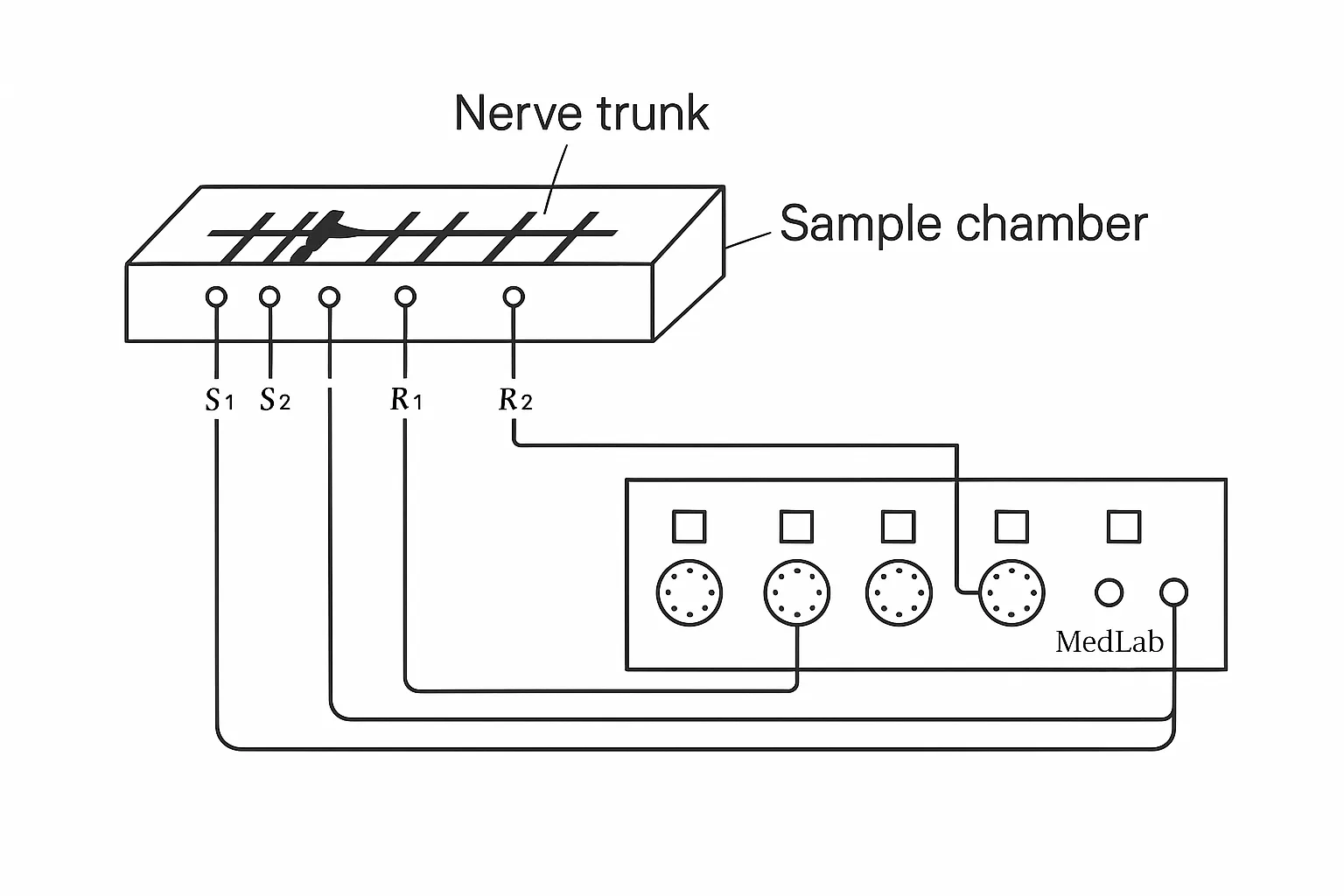
Figure 1 Observing nerve trunk action potential and setup for measuring conduction velocity
2) Line the specimen shield box with filter paper soaked in Ringer solution to maintain humidity and prevent rapid drying of the nerve trunk.
3) Position the nerve trunk specimen on the stimulating electrode, ground electrode, and recording electrodes.
4) Start the ZL-620U medical signal acquisition and processing system and set parameters as appropriate. The stimulation mode can be single or periodic; gradually change the stimulus amplitude.
3. Experimental observations
1) Effect of stimulus intensity on compound action potential: Gradually increase stimulus intensity to find the threshold stimulus that produces a small compound action potential. Continue increasing stimulus intensity; the compound action potential amplitude increases correspondingly until it reaches a maximum and no longer increases with further increases in stimulus intensity. That intensity is the supramaximal stimulus.
2) Carefully observe the biphasic action potential waveform. Record the amplitudes of the positive and negative phases and the total duration at the maximal stimulus.
3) Observe changes in the biphasic waveform when the orientation of the nerve trunk specimen is reversed.
4) Measurement of conduction velocity
- Apply the supramaximal stimulus. In the sampling windows of channels 2 and 4, two biphasic action potentials will be observed sequentially.
- Measure the time from the stimulus artifact to the onset of each action potential: t1 for CH2 and t2 for CH4 (or directly measure the interval between the two action potential onsets). Compute the time difference.
- Measure the distance s between the CH2 and CH4 recording electrode pairs in the specimen shield box (measure from r1 to r2).
5) Observe and measure monophasic action potentials (before damaging the nerve specimen, the refractory-period experiment may be performed).
- Produce a local injury between the two CH4 recording electrodes using forceps or apply a blocking agent; subsequent stimulation will produce a monophasic action potential.
- Record the amplitude and total duration of the monophasic action potential at supramaximal stimulus.
- Compare the rise and fall times of the monophasic waveform and analyze their relationship to the biphasic waveform.
Experimental Results
1) Determine the suprathreshold stimulus range for a specified pulse width, and record the threshold and supramaximal stimulus values.
2) Print biphasic and monophasic action potential waveforms and measure their peak amplitudes and durations.
3) Calculate nerve conduction velocity: v = s / (t2 - t1) (m/s).