ALLPCB


Background
During infection, naive CD8+ T cells are activated, proliferate, and differentiate into effector CD8+ T cells, often accompanied by a rise in body temperature. To determine whether elevated temperature further regulates CD8+ T cell activation and differentiation, it is first necessary to quantify the morphological changes that occur during T cell activation and differentiation.
Amnis' ImageStream high-content imaging flow cytometer combines features of fluorescence microscopy and flow cytometry, enabling rapid analysis of large numbers of cells while acquiring an image for each cell for objective, quantitative analysis. A team at Roswell Park Cancer Institute (RPCI) in New York recently reported that fever-range temperature can enhance immune function, specifically CD8+ T cell function [Thomas A. Mace, JLB, 2011].
Imaging flow cytometry enables quantitative morphology analysis
ImageStream resolved key limitations of conventional flow cytometry and standard imaging: it acquires images for each detected cell while maintaining high-throughput analysis and supports objective, quantitative image analysis. During inflammation or infection, naive CD8+ T cells interact with antigen-presenting cells (APCs) to become activated and differentiate into effector CD8+ T cells. This activation process may coincide with elevated body temperature, but whether temperature elevation further modulates CD8+ T cell activation and differentiation was unclear.
To address this, researchers used ImageStream to quantify morphology and molecular organization during T cell activation and differentiation. They compared mouse CD8+ T cell interactions with APCs after incubation at lower temperatures (33°C and 37°C) and at a higher, fever-range temperature (39.5°C). Their analysis showed that elevated temperature increased the frequency of CD8+ T cell–APC conjugates.
Results: membrane clustering and doublet formation
More importantly, the researchers observed a significant increase in CD8+ T cell numbers in the experimental group. The membrane-associated GM1 and CD8 coreceptor formed clusters, which affected membrane fluidity. These findings suggest that fever-range temperature can influence antigen-specific CD8+ T cell responses by increasing the size of effector cell populations. ImageStream demonstrated robust quantitative image-analysis capability in this study.
The investigators isolated naive CD8+ T cells from mice and incubated them at 33°C, 37°C, or 39.5°C for 6 hours. They then used ImageStream to assess membrane fluidity. Because ImageStream acquires images of detected cells, it can visually reveal whether FITC-labeled GM1 is distributed evenly or clustered on the membrane.
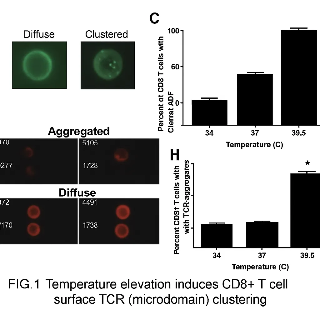
Using image-based quantification across the cell population, the team used the BrightDetail Intensity parameter in the ImageStream software to analyze cells. They found that incubation at 39.5°C significantly increased GM1 clustering on the cell membrane. Prior reports indicate that the TCR signaling complex also clusters on the membrane during T cell activation, so the researchers analyzed that complex as well and observed similar clustering.
To test whether temperature affects formation of CD8+ T cell–APC doublets, naive CD8+ T cells were incubated at 37°C or 39.5°C for 6 hours, then co-cultured with C57BL/6 splenocytes and treated with gp10025-33 peptide in the experimental group. ImageStream was used to quantify the number of conjugates forming immunological synapses.
First, brightfield images and the Aspect Ratio (cell short axis/long axis) were used to gate single cells and doublets (R1 = single cells, R2 = doublets). Then CD8+Thy1.1+ (CD8+ T cells) and CD11b+ (APC) expression was used to gate CD8-APC doublets (Figure 2C). Results showed that higher temperature significantly increased the proportion of CD8+ T cell–APC doublets.
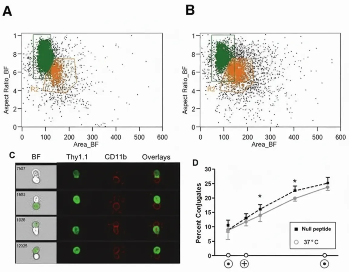
Figure 2. Increased temperature raises the proportion of CD8+ T cell–APC doublets.
Co-receptor clustering and co-localization analysis
The researchers next examined whether CD8 coreceptors also cluster with increasing temperature. The CD8 coreceptor, like TCRβ, is critical for CD8+ T cell activation. Analysis confirmed increased coreceptor clustering at elevated temperature .
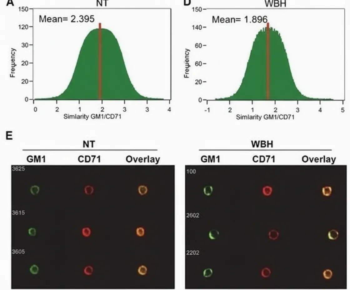
Using the Similarity parameter in the ImageStream software, the team assessed co-localization of CD8 coreceptor and GM1 on the membrane. The similarity values for CD8 and GM1 showed no significant difference before and after temperature elevation (data not shown). To further evaluate GM1 clustering, the investigators analyzed co-localization of GM1 with the negative control CD71. They observed a decrease in similarity for GM1 versus CD71 after temperature elevation (right panels C and D), and images showed that CD71 did not cocluster with GM1 following temperature increase (right panel E).
Conclusion
The study indicates that naive CD8+ T cells and their differentiation into effector cells are temperature-sensitive: exposure to elevated temperature prior to antigen stimulation leads to faster and more effective responses. A major technical bottleneck in such immunology studies is how to obtain statistically meaningful, quantitative data from large numbers of cell images. Conventional flow cytometry lacks morphological information, while fluorescence microscopy typically relies on manual assessment of a limited number of cells. ImageStream high-content imaging flow cytometry overcomes these limitations by enabling rapid, objective quantification of protein translocation, molecular co-localization, morphological changes, endocytosis, and immunological synapse formation.