ALLPCB


Overview of Clinical Applications
Ultrasound medical imaging has developed for more than half a century. Since the 1990s, with rapid advances in medicine, mechanics and materials, computers, and electronic engineering, ultrasound diagnostic instruments have improved in performance, functionality, and application range. Today no hospital can do without ultrasound imaging. Ultrasound offers high spatial resolution, good soft tissue contrast, real-time rapid imaging, simple operation, no contraindications, noninvasive repeatability, portability, and cost-effectiveness. Together with CT, MRI, and nuclear medicine imaging, it forms one of the four essential clinical imaging modalities.
Historical development and technical evolution
Clinical use of ultrasound imaging began in the mid-20th century, initially using A-mode devices to measure thickness of excised organs and explore clinical diagnoses. M-mode was later used to assess cardiac motion in healthy subjects and patients with rheumatic heart disease. In the early 1970s, B-mode two-dimensional imaging that could show organ and lesion morphology was applied clinically, opening the era of transverse and sectional ultrasound imaging. In the mid-1980s, color Doppler ultrasound provided combined information on morphology and hemodynamics, advancing the technique further. The widespread application of computer digital technology in the 1990s enabled successful research into three-dimensional medical ultrasound imaging, bringing ultrasound diagnosis into a new stage of development.
The overall development moved from "point" (A-mode) to "line" (M-mode) to "plane" (2D ultrasound) to "volume" (3D ultrasound); from one-dimensional arrays to two- and three-dimensional arrays; from static imaging to real-time dynamic imaging; from single-parameter diagnosis to multi-parameter diagnosis; and from purely anatomical imaging toward functional, metabolic, receptor/enzyme imaging and molecular imaging integration.
Application of Digital Technology in Ultrasound Diagnostic Equipment
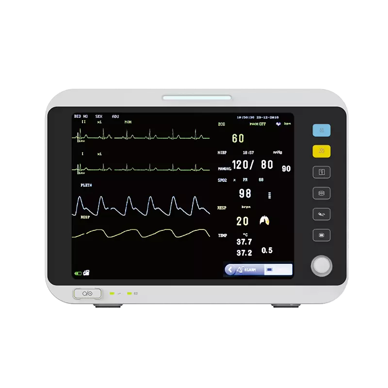
Digitization of ultrasound systems began with digital scan converters and has progressed to fully digital transmit, receive, and imaging chains. Digital techniques are widely adopted in high-performance ultrasound systems, including new probe encoding transmit/receive methods, digital beamforming, digital delay techniques, dynamic receive focusing, dynamic aperture, and dynamic apodization. These technologies have driven higher performance, intelligence, and miniaturization of ultrasound equipment.
Higher-performance systems not only meet diverse clinical diagnostic needs but also support fundamental research and clinical investigations, enabling progress from morphology-focused imaging to physiologic, functional, and molecular imaging. Smart features enable one-button operations that automatically adjust TGC, receive gain, dynamic range, speed scales, Doppler baseline, and other parameters to reduce complex manual adjustments during exams. Miniaturization produces devices with simple form factors, similar to notebook computer size, facilitating bedside, outreach, and emergency applications and expanding clinical use.
With the growth of telecommunications and networks, modern ultrasound systems commonly support the DICOM 3.0 standard interface. DICOM 3.0 covers data dictionaries, information exchange, network communications, media storage and file formats, display and printing, and management functions, and it is extending to cover broader clinical information exchange. This allows ultrasound systems and workstations to integrate with hospital picture archiving and communication systems (PACS) and overall hospital information systems.
Probe Technology Development
The probe, or transducer, is one of the most important components of an ultrasound system; it transmits ultrasound into the body and receives the returning echoes. High-performance, high-quality probes are essential for obtaining high-quality images and enabling new imaging methods. A probe typically consists of a housing, cabling, and the active piezoelectric elements. Piezoelectric materials form the core. Probes evolved from single crystals to multiple elements, and now to arrays with tens, hundreds, or even thousands of elements, increasing the number of array channels.
Current probe development trends include new materials, new manufacturing processes, high-density arrays, higher frequencies, broader bandwidths, and specialty probes. New materials include composite and organic thin-film materials. New processes combine piezoceramics and polymers in defined volume ratios and spatial distributions to produce transducers with high sensitivity, low resistance (for better matching with tissue), and low mechanical quality factors (for broader bandwidth). High-density arrays include 1D arrays (256 elements), 1.5D arrays (e.g., 8×128 elements), and 2D arrays (e.g., 60×60 elements). Frequency ranges: 3 MHz–7 MHz probes are used for abdominal and cardiac diagnosis; 10 MHz–15 MHz probes for superficial organs; 20 MHz–40 MHz probes for ocular and dermatologic imaging; and 100 MHz–200 MHz probes for ultrasound microscopy. Broadband probes have a wide upper and lower operating frequency range, enabling a single probe to transmit and receive different frequencies for shallow-to-deep imaging and supporting frequency compounding, harmonic imaging, and other nonlinear imaging techniques. Specialty probes are shaped for specific uses, such as endoscopic probes for the esophagus, rectum, vagina, urethra, bladder, abdominal cavity, and dedicated vascular probes.
Emerging Imaging Technologies in Ultrasound Diagnosis
1. Three-dimensional ultrasound imaging
Three-dimensional ultrasound imaging represents a significant breakthrough and an emerging clinical modality, providing volumetric information that complements 2D imaging. Based on imaging principles, 3D ultrasound includes static 3D imaging for nonmoving organs and dynamic 3D imaging for moving structures like the heart. Static 3D imaging typically uses a 2D probe with rotational or sector scanning to acquire multiple slice images, which are reconstructed into a 3D volume. Reconstructed images display clear boundaries and surface contours with pronounced depth perception, aiding diagnosis of lesions surrounded by fluid such as hepatic or renal cysts and abscesses, biliary stones and polyps, hydronephrosis, and tumors. Pancreatic and duodenal 3D reconstructions can reveal the three-dimensional anatomy of the pancreatic head and surrounding tissues, assisting diagnosis of lesions affecting the pancreatic head and common bile duct. Vascular 3D reconstructions can generate tree-like images of vessels lacking parenchymal reflection, helping assess vascular course, branching, malformations, and thrombosis. Complex structural lesions, such as facial fetal anomalies and nuchal cord, also show distinctive 3D features. In addition, 3D ultrasound provides spatial location and morphology of tumors, improving localization for ultrasound-guided interventional therapy.
With faster scanning and sampling, adding ECG-synchronized timing to static 3D enables quasi-real-time dynamic 3D imaging (also called four-dimensional imaging). Incorporating velocity information enables real-time 3D imaging (also called five-dimensional imaging). Dynamic 3D imaging can display the origin, position, direction, and spatial relationships of large vessels, detect defects and their morphology and size, and support diagnosis and differential diagnosis of complex congenital heart disease. It can accurately show cardiac geometry and measure function, detect segmental wall motion abnormalities, and provide diagnostic and therapeutic guidance for coronary disease. It also shows valve architecture, aiding diagnosis of stenosis, regurgitation, leaflet clefts, prolapse, and chordae rupture. Dynamic 3D imaging provides volumetric, time-resolved views of intracardiac blood flow to analyze flow direction, regurgitation, and shunting, improving diagnostic accuracy.
2. Wide-field (panoramic) ultrasound imaging
Wide-field ultrasound, also called panoramic or expanded field-of-view imaging, acquires a series of 2D slices while the probe moves and uses software to stitch them into a continuous broad view. The technique provides improved depiction of structural relationships and spatial context, clearly showing lesion location, size, extent, internal echotexture, and adjacency. It enables accurate measurement of large organs and masses and better visualization of tubular structures. Limitations include motion artifacts from tissue or organ movement that can blur images.
Wide-field imaging is widely applied in thoracic and abdominal scanning, obstetrics and gynecology, breast, thyroid, testis, and in musculoskeletal, vascular, and peripheral nerve disease. A single panoramic image can display the entire breast with natural morphology, clear anatomic layers, and distinct lesion characteristics. It can also capture the entire fetus and placenta for assessment in multiple gestations, fetal position, amniotic fluid volume and distribution, and placental location and grading. In musculoskeletal and superficial soft-tissue scanning, high-frequency linear probes can rapidly capture layer-by-layer panoramic anatomy from skin and subcutaneous tissue through muscle, tendon, vessel, peripheral nerve, and periosteum, with clear visualization of normal and pathological features. Wide-field ultrasound has strong development potential and, combined with conventional real-time grayscale and color Doppler imaging, enriches modern ultrasound diagnostics and lays groundwork for ultrasound CT research.
3. Molecular imaging
Molecular imaging uses modern imaging modalities with molecular biology as a foundation to study disease-related pathophysiological and metabolic changes at the molecular level, enabling in vivo visualization of biological processes at cellular and molecular scales. The term emerged in the late 1990s and was formally promoted by the U.S. National Cancer Institute in 1998. Unlike traditional imaging that reveals anatomical consequences of disease, molecular imaging detects cellular and molecular abnormalities that lead to disease, often before structural changes occur. Therefore, molecular imaging can enable earlier detection and assessment of therapeutic effects at the cellular and molecular levels, providing new scientific insight into disease onset, progression, and treatment response.
In ultrasound molecular imaging, targeted microbubble contrast agents labeled with monoclonal antibodies or peptide ligands can be used for targeted diagnostics in cardiovascular disease and oncology, for thrombus and atherosclerotic plaque therapies, and for drug or gene delivery. Microbubbles and other acoustically active agents serve as targeted contrast agents that carry ligands to bind to specific cells for imaging and therapy. Targeted micro- and nano-bubbles have opened a new frontier in molecular imaging. Molecular imaging integrates molecular biology, biochemistry, nanotechnology, genetic engineering, data processing, and image processing, representing a multidisciplinary advancement and an important direction for future medical imaging development.