ALLPCB


Introduction
The emergence of digital imaging technology in the 21st century has enabled improved diagnostic functions, image archiving, and on-demand retrieval. Since digital medical imaging technology appeared in the early 1970s, its importance has grown steadily. Advances in mixed-signal design for semiconductor devices have enabled unprecedented electronic packaging density in imaging systems, driving major advances in medical imaging. At the same time, embedded processors have greatly improved medical image processing and real-time image display, enabling faster and more accurate diagnosis. The convergence of these technologies and emerging electronic health record standards has supported development toward more comprehensive patient care.
This article describes electronic design challenges and recent trends across different imaging modalities, including digital X-ray, magnetic resonance imaging (MRI), and ultrasound systems.
Digital X-ray Systems
Traditional X-ray systems use a film/screen arrangement to detect X-rays transmitted through the body. In a digital X-ray detector system, the signal chain includes a photodetector array that converts radiation into charge, followed by charge-integration circuits and analog-to-digital converters (ADC) to digitize the signal. Figure 1 shows a typical digital X-ray system block diagram.
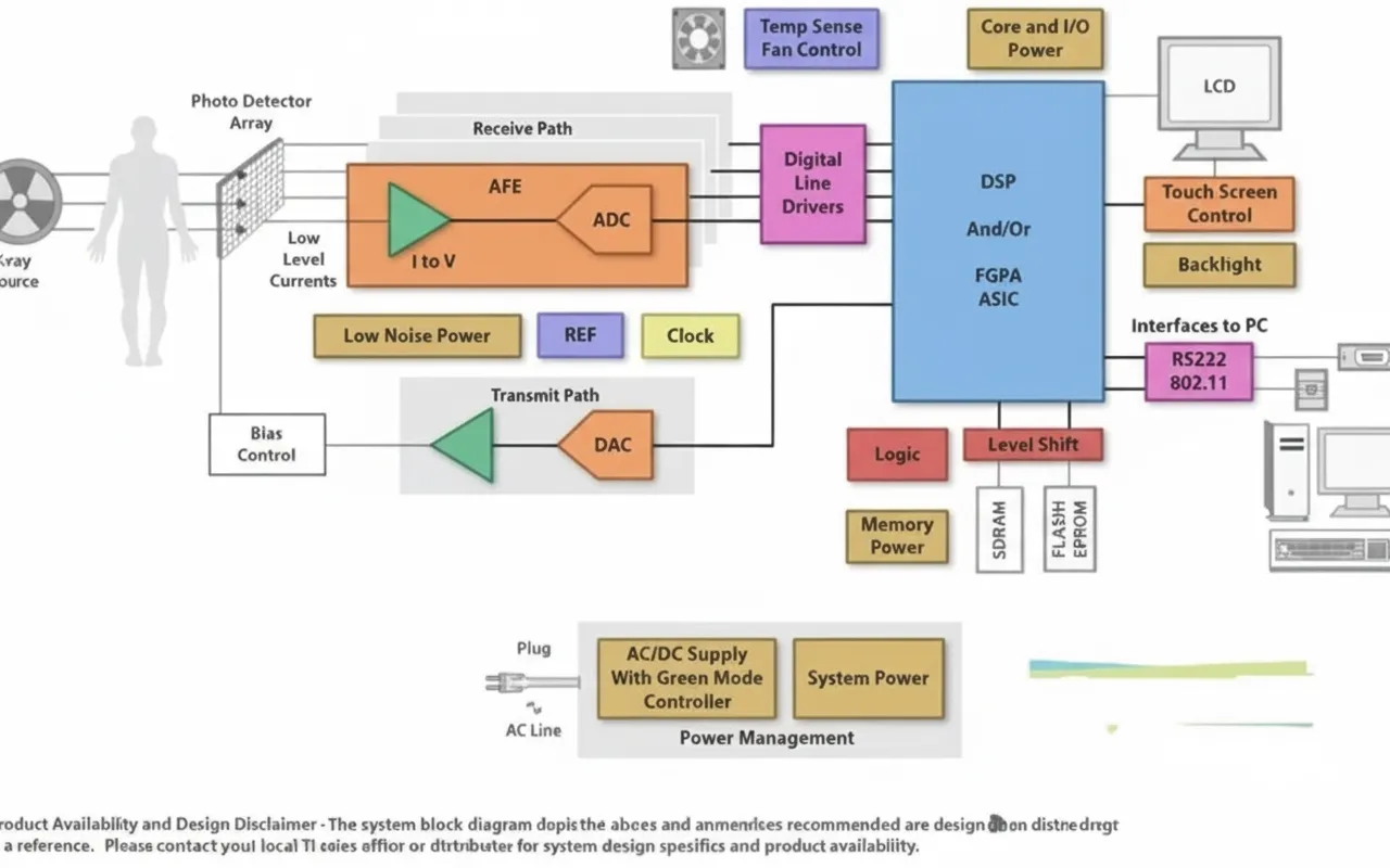
Figure 1 Example block diagram of a digital X-ray system
Digital X-ray system performance is closely tied to the noise performance of the integrators and ADC modules. Achieving high image quality at low power requires a high degree of electronic integration to support the large number of signal channels found in many systems. The combination of high-performance analog components in detector systems and embedded processors that implement advanced image processing functions provides advantages over traditional X-ray systems. This combination supports greater dynamic range, improving image contrast and enabling lower patient X-ray dose, while producing images that can be electronically stored and transmitted.
Ultrasound Systems
The receive-channel signal chain in an ultrasound system typically includes a low-noise amplifier (LNA), variable-gain amplifier (VGA), low-pass filter (LPF), and a high-precision, high-speed ADC. These components are followed by digital beamforming, image and Doppler processing, and other signal-processing software (see Figure 2).
Figure 2 Example block diagram of an ultrasound system
The noise and bandwidth characteristics of the signal-chain components define the upper bound of system performance. At the same time, designers must integrate more high-performance channels into smaller volumes while minimizing power dissipation. A typical handheld ultrasound device may have about 16 to 32 channels, while some high-end systems use 128 or more channels for higher image quality. Reducing the printed circuit board area occupied by all the array channels favors integrating as many channels as possible into the analog front-end IC. Total system power consumption is another key metric for handheld systems. Integrating receive electronics directly into the probe helps shorten the distance between low-voltage analog sources and LNAs, reducing signal loss. Greater integration in the probe also enables more elements for enhanced 3D imaging. Beyond the analog signal-chain considerations, high-performance, low-power embedded processors can perform beamforming and image processing more quickly and efficiently than before.
MRI
Figure 3 shows a typical example of an MRI channel analog signal-processing chain.
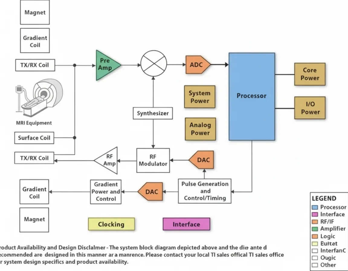
Figure 3 Example block diagram of an MRI system
Whole-body MRI systems may use a coil array with up to about 76 elements or channels. Low-voltage analog inputs are transmitted along long coaxial cables from limb coils to the front-end preamplifier. Two key requirements arise for MRI receive chains: how to achieve high signal-to-noise ratio (SNR) on the order of 84 dB (roughly 14 bits), and how to realize a very high overall system dynamic range, on the order of 150 dB/Hz. Achieving high SNR requires a very low-noise, high-performance front-end amplifier. Techniques such as dynamic gain adjustment or analog input compression can help meet the dynamic-range requirement.
Increasing the number of coils can improve image coverage and reduce scan time. More coils may require optimized communication between coils and preamplifiers, and the use of high-speed digital or optical links places additional demands on the main system. Higher integration could change current system partitioning and place electronics closer to the coils. This may require non-magnetic semiconductor packaging and stricter power and area constraints. Successfully meeting these requirements reduces input signal attenuation and yields higher-quality medical images.
Conclusion
Digital imaging remains one of the most active technology development areas in medical electronics. Advances in IC analog/mixed-signal functionality and embedded processing continue to drive improvements in imaging-system performance. These technological developments improve imaging capability and the quality of diagnostic and clinical care.