ALLPCB


The emergence of digital imaging in the 21st century has enabled improved diagnostic capability, electronic image archiving, and anytime-anywhere retrieval. Since digital medical imaging first appeared in the early 1970s, its importance has grown. Advances in semiconductor devices and mixed-signal design have enabled unprecedented electronic packaging density in imaging systems, contributing to major advances in medical imaging. At the same time, embedded processors have greatly increased the capability for medical image processing and real-time image display, enabling faster and more accurate diagnosis. The integration of these technologies and the adoption of electronic health record standards have supported improved patient care.
This article reviews electronic-design challenges and recent developments across different imaging modalities, including digital X-ray, MRI, and ultrasound systems.
Digital X-ray Systems
Traditional X-ray systems use a film/screen assembly to detect X-rays transmitted through the body. In detector-based digital X-ray systems, the signal chain includes a photodetector array that converts radiation into charge. That is followed by charge-integrator circuits and analog-to-digital converter (ADC) circuits to digitize the input. Figure 1 shows an example block diagram of a typical digital X-ray system.

Figure 1. Example digital X-ray system block diagram
Digital X-ray performance is closely linked to the noise performance of integrators and ADC modules. To achieve high image quality at low power, the electronic integration required to support large numbers of signal channels sets demanding technical requirements. The combination of many high-performance analog components in the detector subsystem and embedded processors performing advanced image processing gives detector-based X-ray systems several advantages over conventional film-based systems. This combination supports larger dynamic range, enabling improved image contrast and lower patient X-ray exposure, while producing digitally storable and transmittable images.
Ultrasound Systems
The receive-channel signal chain in ultrasound systems typically includes a low-noise amplifier (LNA), variable-gain amplifier (VGA), low-pass filter (LPF), and high-speed high-precision ADC. These front-end components are followed by digital beamforming, image and Doppler processing, and other signal-processing software (see Figure 2).
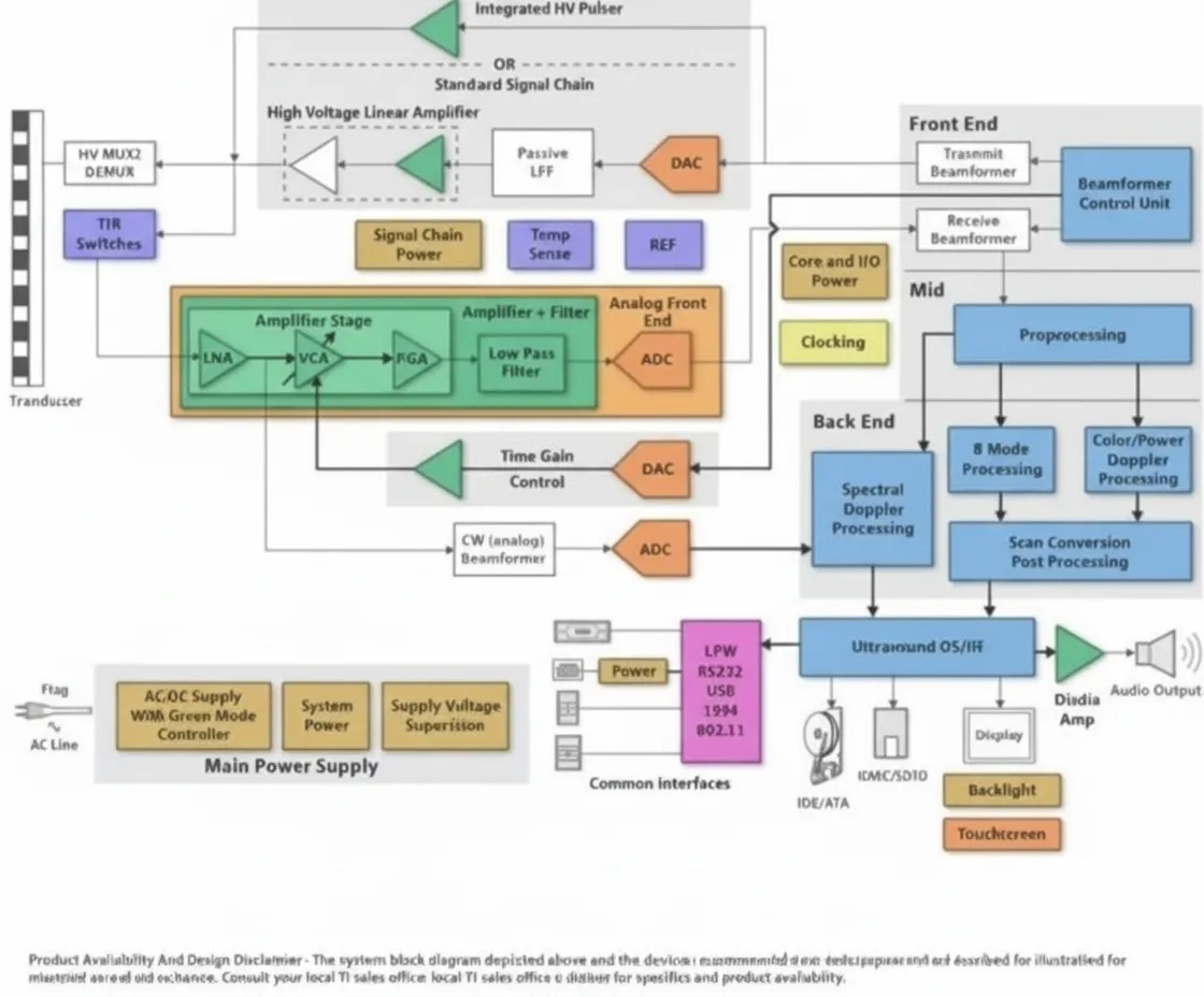
Figure 2. Example ultrasound system block diagram
The noise and bandwidth characteristics of the signal-chain components define the upper limit of system performance. In addition, there is pressure to integrate more high-performance channels into a smaller area while reducing overall system power. A typical handheld ultrasound system may have roughly 16 to 32 channels, while some high-end systems exceed 128 channels to achieve higher image quality. To reduce PCB area consumed by all these array channels, emphasis is placed on integrating as many channels as possible into the analog front-end ICs. Total system power is another important metric for handheld devices. Integrating the receive electronics directly into the probe is another innovation.
Placing electronics in the probe shortens the distance between the low-voltage analog signal sources and the LNAs, reducing signal loss. Integration also increases the number of elements in the probe, supporting improved 3D imaging. Beyond these analog signal-chain considerations, high-performance, low-power embedded processors now perform beamforming and image processing tasks in portable devices faster and more efficiently than before.
MRI
Figure 3 shows an example of a typical MRI channel analog signal-processing chain.
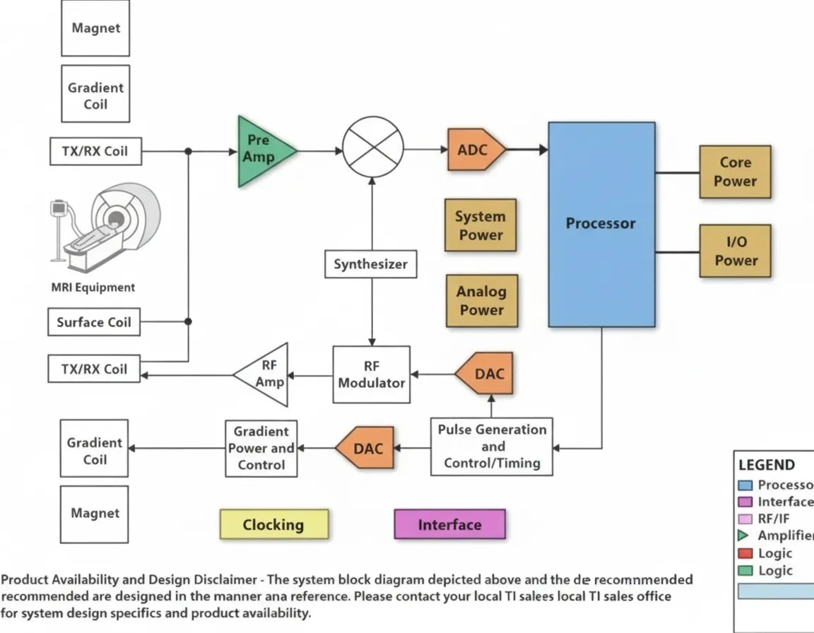
Figure 3. Example MRI system block diagram
Whole-body MRI systems may use coil arrays with up to about 76 elements or channels. Low-voltage analog inputs travel along long coaxial cables from limb coils to the front-end amplifiers. Two key requirements for MRI receive chains are achieving high signal-to-noise ratio (SNR) — typically on the order of 84 dB (about 14 bits) or better — and attaining very large overall system dynamic range (on the order of 150 dB/Hz). Achieving high SNR requires a very low-noise front-end amplifier. Techniques such as dynamic gain control or analog input compression can help meet the high dynamic-range requirement.
Increasing the number of coils in MRI systems improves image coverage and can reduce scan time. More coils may require further optimization of the signal link between coils and front-end amplifiers, and the use of high-speed digital or optical links may require additional system-level optimization. Higher integration can shift system partitioning, placing electronics closer to the coils. That can require non-magnetic semiconductor packaging and stricter power and area constraints. Successfully meeting these requirements reduces input-signal attenuation and improves image quality.
Conclusion
Digital imaging remains one of the most active areas of technology development in modern medical imaging. Advances in IC analog/mixed-signal capabilities and embedded processing continue to drive performance improvements in imaging systems, which in turn enhance diagnostic and patient-care quality.