ALLPCB


Project overview
1.1 Introduction
Blood oxygen saturation (SaO2) is a physiological parameter that reflects the blood's oxygen-carrying capacity and is important in clinical diagnosis. Measurement methods are invasive or noninvasive. The invasive method requires blood sampling followed by electrochemical analysis with a blood gas analyzer to obtain partial pressure of oxygen (PO2) and compute SaO2. This method is cumbersome and cannot provide continuous monitoring. Noninvasive measurement mainly uses a dual-beam transmission method, estimating SaO2 from the ratio of the relative changes in absorbance when red and infrared light of different wavelengths pass through biological tissue.
Because biological tissue is a strongly scattering, weakly absorbing, anisotropic optical system that does not fully conform to the classical Beer-Lambert law, the relationship between the measured ratio of relative absorbance changes for red and infrared light (the R/IR ratio) and arterial oxygen saturation (SaO2) can only be modeled approximately. In addition, nonidealities of the internal light sources in noninvasive pulse oximeters introduce errors that generally make noninvasive measurements less accurate than invasive methods. This design examines these issues and proposes a portable noninvasive pulse oximeter architecture intended to improve SaO2 measurement accuracy.
1.2 Background and motivation
Adequate oxygen supply is fundamental to life, and arterial oxygen saturation is a key indicator of oxygenation; its measurement is important in clinical practice. Although the Van Slyke method provides precise measurements on blood samples, it cannot perform continuous monitoring and involves a complicated process that is inconvenient for clinical staff and uncomfortable for patients. Pulse oximetry provides noninvasive, continuous, fast monitoring of arterial oxygen saturation (SaO2) and has been adopted in clinical settings, particularly for monitoring critically ill patients during surgery. Pulse oximeters are widely used in operating rooms, recovery rooms, emergency wards, and for research in exercise and sleep studies.
AVR32-series microcontrollers offer low power consumption and good analog performance. Their use in single-chip portable medical devices can minimize the number of external components. In this design, power consumption can be further reduced by controlling LED on-time and operating frequency, enabling an ultra-low-power system suitable for portable applications.
Requirements analysis
2.1 Functional requirements
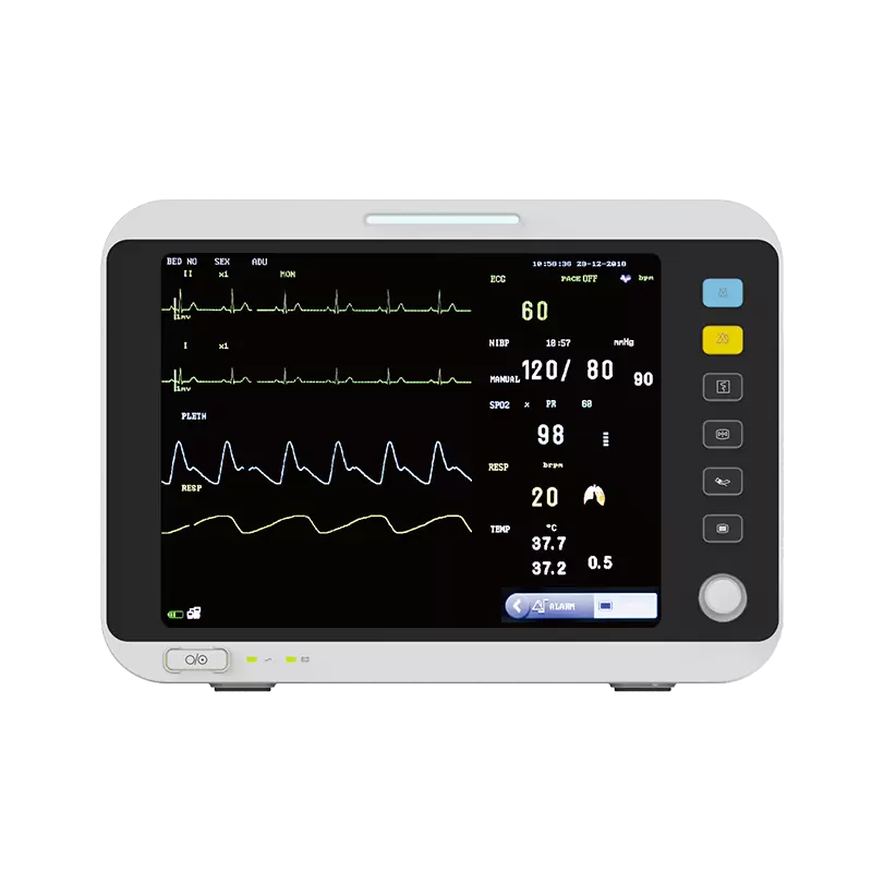
The system acquires SaO2 and heart rate via an external optical probe and displays results on an LCD. The optical probe contains two LEDs: one visible red LED and one infrared LED. The probe can be placed on the fingertip or earlobe. After the two LED beams pass through tissue, the MCU detects changes in the two light intensities to compute blood oxygenation. When blood oxygen saturation or heart rate falls below preset thresholds, the oximeter issues an alarm signal to notify the user.
2.2 Performance requirements
- Measurement method: dual-wavelength measurement;
- Numeric display: large-screen LED or LCD display; SaO2 and pulse values clearly visible and not limited by ambient lighting, suitable for emergency settings;
- Pulse display: three-color analysis system for quick indication of pulse quality;
- Measurement range: SaO2: 0–100%; heart rate: 18–300 beats per minute;
- Measurement accuracy: SaO2: ±1 S.D.; heart rate: ±3%;
- Probe compatibility: suitable for adult, pediatric, and neonatal probes;
- Data storage and fast, responsive SaO2 reporting; suitable for sleep analysis and able to communicate with a PC via USB;
- Additional display: current time and temperature;
- Power: battery operation using six AA cells, continuous operation over 24 hours.