ALLPCB


Technological advances in medical imaging during the last century created unprecedented opportunities for noninvasive diagnosis and established medical imaging as an integral part of healthcare systems. One of the principal innovation areas representing these advances is the interdisciplinary field of medical image processing.
This rapidly evolving field spans the workflow from raw data acquisition to digital image transmission, forming the basis of the complete data flow in modern medical imaging systems. Today, these systems provide increasingly higher resolution in spatial and intensity dimensions and faster acquisition times, producing large volumes of high-quality raw image data that must be correctly processed and interpreted to yield accurate diagnoses.
This article focuses on the key areas of medical image processing, considers the context of specific imaging modalities, and discusses the main challenges and trends in the field.
Core areas of medical image processing
Many concepts and methods are used to structure the field of medical image processing, each emphasizing different core areas. These aspects form three main processes in the field: image formation, image computation, and image management.
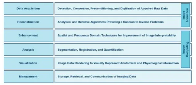
Figure 1. Structural classification of topic types in medical image processing.
The image formation process comprises data acquisition and image reconstruction steps that address mathematical inverse problems. The purpose of image computation is to improve the interpretability of reconstructed images and extract clinically relevant information. Finally, image management handles compression, archiving, retrieval, and transmission of acquired images and derived information.
Image formation
Data acquisition
The first required step in image formation is acquiring raw imaging data. This data contains the raw information that describes the physical quantities captured from the body's internal structures. These data form the basis for all subsequent image-processing steps.
Different imaging modalities exploit different physical principles and therefore involve detection of different physical quantities. For example, in digital radiography (DR) or computed tomography (CT) it is the incident photon energy; in positron emission tomography (PET) it is photon energy and detection timing; in magnetic resonance imaging (MRI) it is parameters of the radiofrequency signals emitted by excited atoms; and in ultrasound it is echo parameters.
Regardless of modality, the data acquisition process can be subdivided into detection of the physical quantity, conversion of that quantity to electrical signals, preconditioning of the acquired signals, and digitization. A general block diagram illustrating these steps, which applies to most medical imaging modalities, is shown in Figure 2.

Figure 2. General block diagram of the data acquisition process.
Image reconstruction
Image reconstruction is the mathematical process that forms images from acquired raw data. For multidimensional imaging, it can also involve combining multiple datasets captured at different angles or time steps. This part of medical image processing addresses inverse problems, which are a fundamental topic in the field. Algorithms used to solve these problems fall mainly into two categories: analytical and iterative.
Typical analytical methods include filtered back projection (FBP), widely used in tomography; the Fourier transform (FT), especially relevant in MRI; and delay-and-sum (DAS) beamforming, a standard technique in ultrasound. These algorithms are computationally efficient and well suited to the processing power and time constraints of many systems.
However, analytical methods rely on idealized models and have limitations, such as difficulty handling complex factors like the statistical nature of measurement noise and detailed system physics.
Iterative algorithms overcome many of these limitations, offering greater robustness to noise and the ability to reconstruct optimal images from incomplete raw data. Iterative methods typically employ system and noise statistical models. Starting from an initial object estimate, they compute projected data using model parameters and compare these projections to the acquired data; the difference defines updates to the object estimate. This process repeats for multiple iterations until a cost function measuring the discrepancy between the estimate and the true object is minimized. Common iterative approaches include maximum likelihood expectation maximization (MLEM), maximum a posteriori (MAP) methods, algebraic reconstruction techniques (ARC), and many other variants widely applied across imaging modalities.
Image computation
Image computation covers the computational and mathematical methods applied to reconstructed image data to extract clinically relevant information. These methods are used for enhancement, analysis, and visualization of imaging results.
Enhancement
Image enhancement optimizes transformed representations of images to improve interpretability. Methods are broadly classified into spatial-domain and frequency-domain techniques.
Spatial-domain techniques operate directly on image pixels and are particularly useful for contrast optimization. These techniques often rely on logarithmic, histogram, and power-law transforms. Frequency-domain methods use frequency transforms and are well suited for smoothing and sharpening by applying different types of filters.
These methods reduce noise and inhomogeneity, optimize contrast, enhance edges, remove artifacts, and improve other characteristics that are critical for accurate subsequent image analysis and interpretation.
Analysis
Image analysis is a core process within image computation, and its methods fall into three main categories: image segmentation, image registration, and image quantification.
Segmentation divides an image into meaningful contours representing different anatomical structures. Registration ensures multiple images are correctly aligned, which is essential when analyzing temporal changes or combining images from different modalities. Quantification determines properties of identified structures such as volume, diameter, composition, and other anatomical or physiological information. These processes directly affect the quality of image-based assessments and the accuracy of clinical outcomes.
Visualization
Visualization presents image data in intuitive formats that represent anatomical and physiological information along defined dimensions. Interactive visualization can assist early and intermediate stages of image analysis, for example by supporting segmentation and registration, and can present final optimized results.
Image management
The final component of medical image processing involves managing acquired information, including technologies for image storage, retrieval, and transmission. Several standards and technologies address different aspects of image management. For example, picture archiving and communication systems (PACS) provide economical storage and access to images from multiple modalities, and the Digital Imaging and Communications in Medicine (DICOM) standard is used for storing and transmitting medical images. Techniques for image compression and streaming efficiently support these tasks.
Challenges and trends
Medical imaging is a relatively conservative field in which translation from research to clinical application can take a decade or more. Its complexity creates multiple challenges across the scientific disciplines that compose it, which in turn drive steady development of new methods. These developments represent the main trends in current medical image processing.
The data acquisition domain benefits from innovative hardware developed to improve raw data quality and enrich its information content. Integrated front-end solutions enable faster scan times, finer resolution, and advanced architectures such as combined ultrasound/mammography, CT/PET, or PET/MRI systems.
Fast and efficient iterative reconstruction algorithms are increasingly replacing analytical methods. They can significantly improve PET image quality, reduce CT radiation dose, and enable compressed sensing in MRI. Data-driven signal models are replacing hand-crafted models, offering better solutions to inverse problems from limited or noisy data. Key research areas in image reconstruction include system physics modeling, development of signal models, optimization algorithms, and image-quality assessment methods.
As imaging hardware captures larger datasets, algorithms become more complex and demand more efficient computation. This challenge is being addressed by more powerful graphics processors and multiprocessing techniques, which open new opportunities for translating research into practice.
The main trends and challenges related to image computation and management span many topics, examples of which are shown in Figure 3.
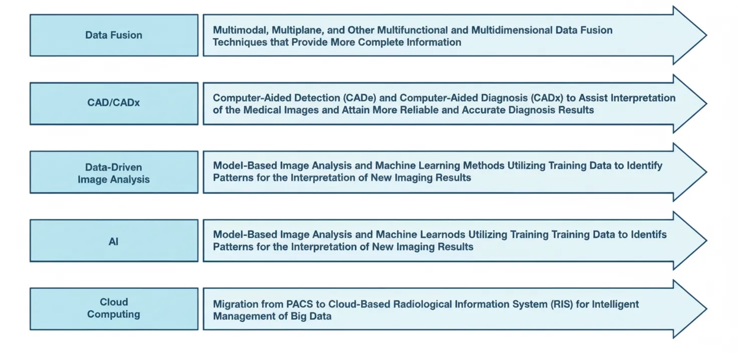
Figure 3. Examples of major trend topics in contemporary medical image computation.
Ongoing development of new techniques is narrowing the gap between research and clinical application, facilitating integration of medical image processing into physician workflows and helping to achieve more accurate and reliable imaging results.
Conclusion
Medical image processing is a complex interdisciplinary field spanning mathematics, computer science, physics, and medicine. This article outlines a simplified but structured framework of the field, highlighting its main topics, trends, and challenges. Data acquisition is one of the first and most important areas, as it defines the initial quality level of raw data used in all subsequent stages of the medical image processing workflow.