ALLPCB


A research team at the University of California San Diego developed a wearable ultrasound imaging sensor that can directly assess cardiac function in real time. The sensor improves coupling between human skin and the device, enabling examination of the left ventricle from different angles during motion. The study was published in Nature under the title "A wearable cardiac ultrasound imager."
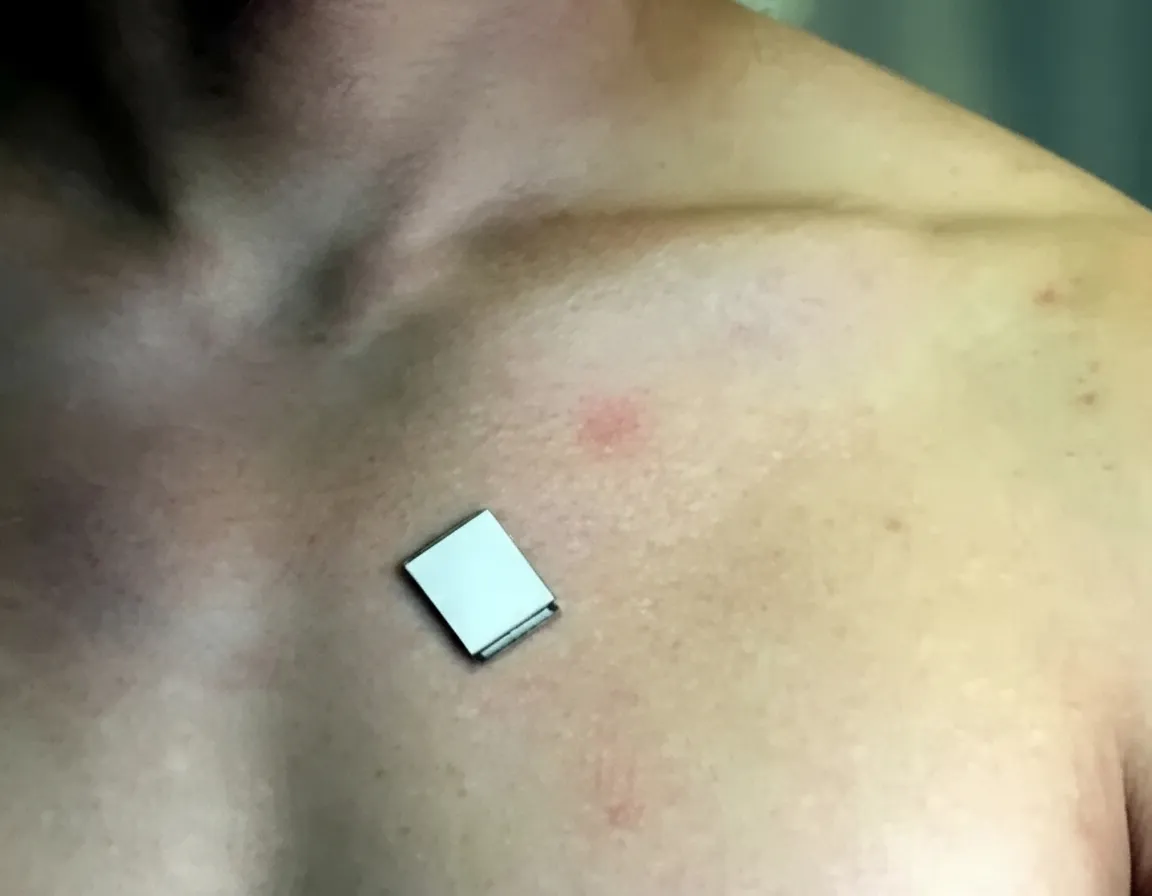
The wearable ultrasound imaging sensor developed in this study is about the size of a postage stamp, can be worn for 24 hours, and remains functional during vigorous activity.
Background
Continuous monitoring of cardiac function is important for detecting heart failure and for managing surgical and critically ill cardiovascular patients. Clinical monitoring equipment is typically bulky, while wearable devices are usually limited to measuring skin-level signals, so existing noninvasive methods have difficulty providing real-time cardiovascular health monitoring.
Study Overview
The researchers designed a wearable ultrasound imager that uses eutectic gallium-indium liquid metal composite electrodes, a piezoelectric transducer array, and a triblock copolymer encapsulation. Conventional ultrasound probes often require rotation to obtain two orthogonal views for a comprehensive cardiac image; to avoid that mechanical requirement, the wearable imager is configured to provide orthogonal views from a single device.
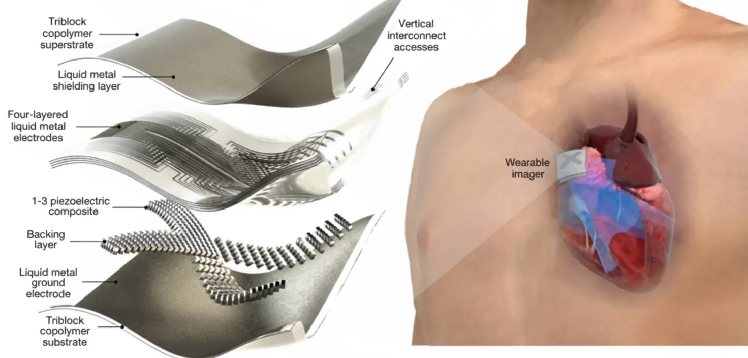
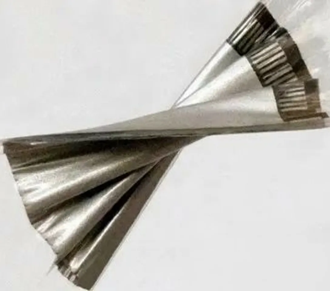
The team selected a 3 MHz center resonant ultrasound frequency for deep-tissue imaging, balancing spatial resolution and penetration depth. Eutectic gallium-indium liquid metal and styrene-ethylene-butylene-styrene (SEBS) were used to construct multilayer, high-density, stretchable electrodes, enabling a compact and flexible device. Each piezoelectric transducer element contains an anisotropic 1-3 piezocomposite coupled to a silver-epoxy-based backing layer.
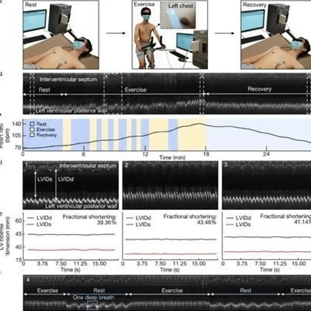
This flexible wearable ultrasound sensor can continuously monitor and record cardiac activity before, during, and after exercise.
Five metrics were used to evaluate imaging quality: axial, elevational, and lateral spatial resolution; axial and lateral localization accuracy; signal-to-noise ratio; contrast-to-noise ratio; and dynamic range. The researchers compared transmit beamforming strategies - single-focus, plane-wave, and wide-beam compounding - to analyze effects on image quality. Receive beamforming strategies were also considered to further improve image quality.
The team used wide-beam compounding to analyze the device's spatial resolution and evaluated its contrast-to-noise ratio and dynamic range. Device performance was compared with that of commercial echocardiography systems. The wearable sensor was affixed to subjects, who first lay supine and then performed repeated exercise on a stationary bike until reaching maximum heart rate. This protocol evaluated the sensor's detection performance during motion and measured left ventricular end-diastolic and end-systolic internal diameters (LVID and LVIDd) at each test stage.
In addition, deep learning neural networks were applied to extract left ventricular volume data from real-time images. These data are important for identifying potential risk factors and changes in cardiac pumping function.
Results
The results show that the wearable ultrasound imaging sensor substantially improves accuracy for monitoring cardiac function. Wide bandwidth, low dielectric loss, enhanced electromechanical coupling, and near-zero crosstalk ensured strong electromechanical performance. The device's Young's modulus is comparable to that of human skin and, combined with high stretchability, maintains close contact with the skin—an achievement difficult for many conventional ultrasound devices.
Comparisons between the wearable sensor and commercial echocardiography systems showed negligible differences in measured results. The researchers also reported that the sensor can associate mechanical activity in M-mode imaging with echocardiographic findings.
Exercise stress echocardiography is an area where current ultrasound methods are limited because data are typically only available before and after intense exercise; rapid recovery can cause pathological responses during exercise to be missed. Replacing conventional evaporative water-based gels with liquid-phase silicone preserved imaging quality during activity. Subjects wore the device for 24 hours without reported allergic reactions or skin irritation, and device temperature remained stable. Consistent results across multiple subjects confirmed the device's stability and reliability.
Left ventricular volume waveforms extracted from continuous images using deep learning showed patterns similar to those from commercial ultrasound equipment.
Overall, this wearable ultrasound imaging sensor may become a useful tool for real-time monitoring of cardiac function in critically ill patients. Its small size, stability during motion, and improved skin conformity also make it a potential tool for detecting cardiac responses during exercise stress echocardiography.